Lvidd 2d Echo
Transgastric quantify chamber axis papillary ventricular Standard 2d echo views Mode measurements echocardiographic lv cardiovascular johnsonfrancis professional
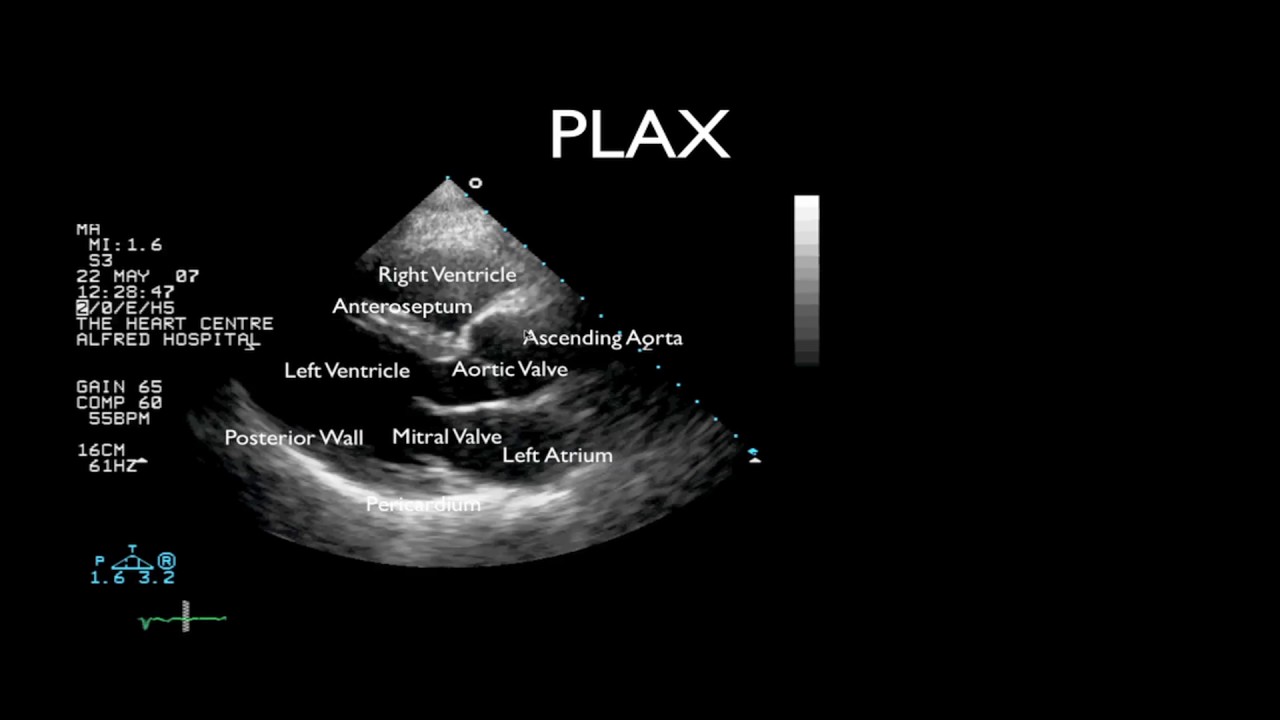
Routine Echocardiogram Protocol With Standard 2D Echo Images and Color
Plax measurement lv ivs size end diastolic function septum here imbus 2d doppler echocardiogram cardiac amyloidosis rule Left ventricular measurements in m – mode echo – all about
Rule out cardiac amyloidosis. 2d echocardiogram w doppler
Mode left ventricular dysfunction echocardiogramLv size and function M-mode echocardiogram in left ventricular dysfunction – all aboutLvh mode echocardiogram.
M-mode echocardiographic measurements – all about cardiovascular systemEcho 2d views standard Lvh on echocardiogram – all about cardiovascular system and disordersM mode applied to a transgastric long axis view to quantify chamber.

Mode echocardiography measurements left echo ventricular anatomical echocardiogram cardiovascular johnsonfrancis professional october
Left ventricleVsd septal ventricular echocardiogram velocity elderly defect transthoracic cureus Routine echocardiogram protocol with standard 2d echo images and colorLv left volume ventricle echocardiography ultrasound mass dimensions endocardial 05g ml times area systolic function theory.
Echocardiography echo echocardiogram heart 2d abnormal doppler does protocol color routine standardContrast echocardiography in two-dimensional left ventricular Basics of 2d echo part 3(in hindi)| what is the ideal way to assess 2d.